Solution to create 3D anatomical models using 3D scanning technology helps to digitize real organs, reverse engineer and 3D print for medical teaching. 3D Master provides professional 3D scanning services to help training units reduce costs, shorten time and improve the quality of anatomy.
In the medical field, high-quality anatomical models play an important role in teaching and research. However, traditional plastic models or 2D image simulations cannot show the detail, size and real shape of internal organs. Therefore, the solution to create anatomical models using 3D scanning technology is becoming an inevitable trend.
3D scanning technology allows accurate data collection from real organ models or available sample models. This data is then reverse engineered into a complete 3D model that can be 3D printed for study and research. This is a big step forward for the medical industry to access more intuitive, modern and accurate training methods.
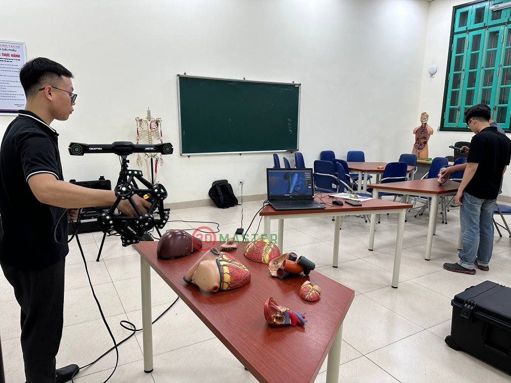
3D Master scan 3D medical learning tools with Creaform Metrascan
3D scanning is a method of digitizing objects by using light or laser to record the entire shape, size, curvature and small details of the object. The result is a high-resolution point cloud, thereby creating a 3D model accurate to the micrometer.
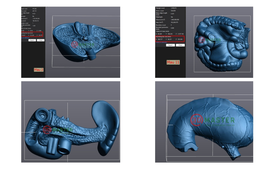
3D scanning results of medical learning tools
In anatomy, 3D scanning offers a superior advantage because it can faithfully reproduce each anatomical structure, from soft tissue to hard tissue. This makes it easy for students and doctors to observe, analyze and research without having to rely on real samples that are difficult to preserve and limited in quantity.
The use of 3D scanning in medicine brings many outstanding applications. One of the most popular applications is creating 3D organ models from real samples for teaching purposes. These models can be 3D printed with biological materials or engineering plastics, meeting the requirements of simulating medical structures.
In addition, 3D scanning also supports tissue deformation analysis, comparison between different organ samples or assessment of differences during the research process. For medical schools, 3D anatomical models created from 3D scanning help students experience a more intuitive and accessible training method than static images.
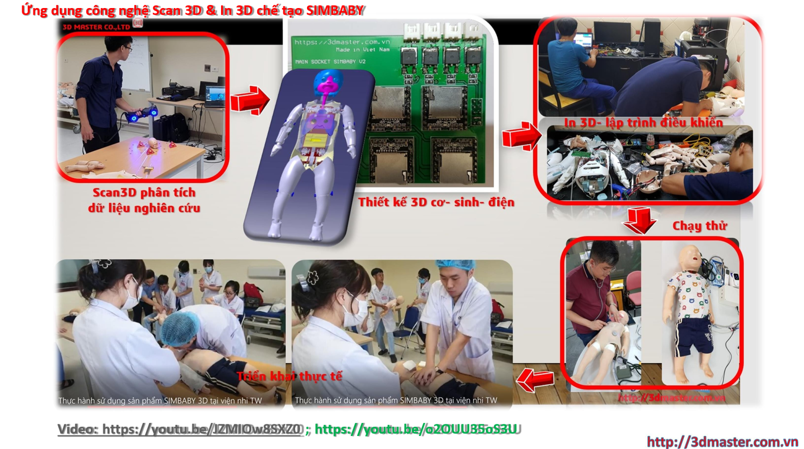
Applying 3D Scanning & 3D Printing Technology to Make SIMBABY
Reverse engineering from 3D scanning data helps ensure that the 3D model retains the true shape of the internal organs. Thanks to that, the 3D printed model meets strict requirements for accuracy. Reverse engineering also opens up the ability to customize the model according to research needs such as zooming, separating layers, adding annotations or simulating pathology. This is an advantage that traditional anatomical models cannot meet.
After completing the reverse engineering, the 3D model is transferred to the 3D printing system. Current 3D printing technology can reproduce the color, texture and shape of internal organs realistically. This makes the learning and research process easier to understand, intuitive and vivid
3D printing from 3D scan data also helps save costs significantly because it can create many copies of the model to serve many different classes.
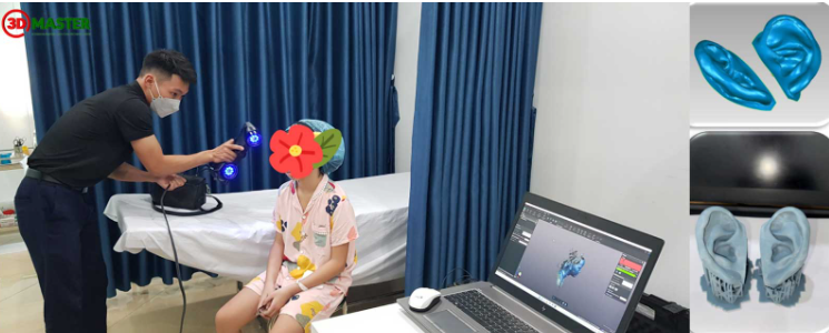
3D scanner application to create 3D models for medical examination and treatment
Many medical universities, training centers and research institutes have switched to using 3D scanning solutions thanks to their outstanding advantages. High accuracy, the ability to reproduce anatomical details and flexibility in design are important factors that make this method popular.
In addition, 3D scanning creates a digital data source that can be stored for a long time to serve many different studies in the future. This is the foundation for the modern medical digitalization trend.
3DMaster is a leading unit in the field of 3D scanning and reverse engineering. With a modern 3D scanning system, a team of experienced engineers and standard processes, 3DMaster meets all requirements for creating high-quality 3D anatomical models.
The unit has successfully implemented many projects of 3D scanning of internal organ models, reverse engineering for teaching and research for medical schools and large training centers across the country. Powerful data processing capacity helps shorten implementation time while still ensuring absolute accuracy.
3D scanning technology is revolutionizing the way anatomical models are created and used in medicine. Thanks to the ability to accurately recreate real organs, combining reverse engineering and 3D printing, this solution helps to increase the efficiency of teaching and research to a new level. In the context of rapidly developing medical digitalization, 3D scanning will certainly continue to play a key role.
If you need 3D scanning of organ models, reverse engineering or 3D printing for teaching and research, contact 3DMaster today. We provide professional 3D scanning services, fast speed, high accuracy and suitable for all medical training facilities.
Hotline - Zalo - LINE - Telegram - WhatsApp - Viber - Kakaotalk: +84 982 089 198 | 0986333960
Email: cuong3dmaster@gmail.com | hung3dmaster@gmail.com | tech3dmaster@gmail.com
Website: https://3dmaster.com.vn